Procedure
Salisphere Formation
This is an in vitro (controlled environment) culture system. To learn more, visit our Further Research page.
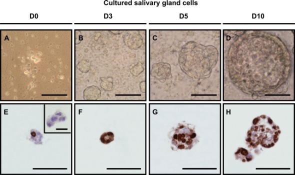
A shows the disassociation of a mouse's submandibular gland in a cell suspension. The first evidence of the salispheres are in B as a result of proliferation, where they grow in C and D. E-H shows the cells in the culture actively dividing through staining of the cells.
The salispheres were found to have originated from duct cells. A salisphere differentiates into the different salivary gland cell types within a sphere by coming into contact with similar cell types. The c-Kit expressing stem cell pool came in contact with the cells from the original dissassociation and was able to produce more cells. Consequently, the amount of stem cells expressing c-Kit decreased.
Salispheres Containing Stem Cells
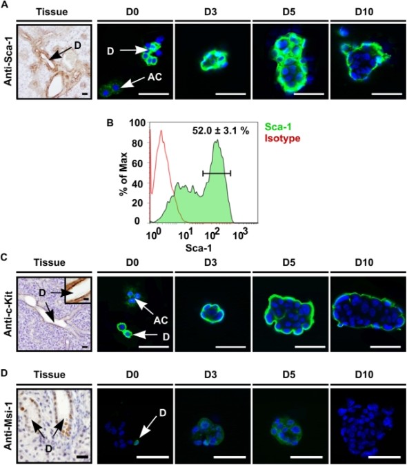
The culture tested for the presence of three different yet common stem cell markers: Sca-1, c-Kit, and Musashi-1 using specific antibodies. In A, Sca-1 was present in the initial gland and the beginning of the culture, however it was not expressed in the acinar cells after 10 days. In C, c-Kit was only expressed in the excretory duct cells in the tissue but was expressed similarly to Sca-1, disappearing after a few days. Musashi-1, in D, also diminished.